Streptococcus (first part)
Hi, after long time,
Today, we will explain Streptococcus
with its structure and classification, so let’s begin:
---------------------------------------------------------------------------------------------------------------------
*Pyogenes
cocci are those in which the hallmarks (special characteristic) of the
infective process results in formation of pus. And they are classified into Gram(+ve)
pyogenes such as Streptococcus and staphylococcus tend to cause
diseases by exotoxins while Gram (-ve) pyogenes such as Neisseria
and Haemophilus causes diseases depending on host’s response to lipopolysaccharides
(LPS) (endotoxin) as LPS cause releasing of inflammatory cytokines. *
Streptococcus are aerobic and facultative anaerobic Gram-positive cocci, arranged in pairs (diplococci) such as Streptococcus pneumonia or chains such as Streptococcus pyogenes.
They are nonmotile, non-sporing,
have hyaluronic acid capsules and catalase negative
by which they are distinguished from staphylococci.
They are classified based
on:
1) Hemolysis in blood
agar (bacteriologically):
Brown classified these aerobic streptococci into three groups on the
basis of their growth in 5% horse blood agar:
a)
Alpha-hemolytic streptococci: These
cocci produce colonies surrounded by a narrow zone (greenish zone) of hemolysis
with persistence of some partially lysed RBCs such as Virdians streptococci
and Streptococci Pneumonia.
b)
Beta-hemolytic streptococci: These
cocci produce a clear, colorless zone of hemolysis around the colonies such as Group
A (S. pyogenes), Group B (S. agalactiae), Group C (S. equismilis, S. equi, S. zooepidemicus)
and Group D.
c)
Gamma-hemolytic streptococci: These
do not produce any hemolysis or discoloration on blood agar such as Enterococcus
and Non-enterococcus.
2) Antigenic structure: (immunologically):
Lancefield classification is a serological classification of beta-hemolytic
streptococci. This classification is based on detection of group-specific
carbohydrate antigen (C-antigen) (it will be explained later) on the cell
wall.
Beta-hemolytic streptococci are classified
into 21 serological groups (A to W, except I and J).
Note: S.viridans and
S. pneumonia don’t possess C- carbohydrate thus
not serogrouped.
Based on the M, T, and R
protein antigens ((it will be explained later) present on the cell
surface, S. pyogenes have been further classified into 80 serotypes. This
classification is known as Griffith typing.
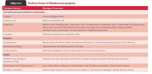
Structure of Streptococcus:
We will talk about cell wall
components and antigenic structure in addition to virulence factors, So
focus!!!!!
First: cell wall
components and antigenic:
1)
Group-Specific Carbohydrate:
The cell wall contains a
group-specific polysaccharide that forms approximately 10% of the dry weight of
the cell. It is a polymer of N-acetylglucosamine and rhamnose.
2)
Type-Specific Protein:
The cell wall of S. pyogenes has
three major proteins, M, T, and R proteins.
a)
M proteins: is the important protein and is considered chief virulence factor
of cocci as it inhibits phagocytosis and facilitates attachment of
cocci to epithelial cells.
M protein is alpha helix
consisting of carboxyl terminus and amino terminus. This carboxyl
terminus is attached to cytoplasmic membrane and is highly conserved
while amino terminus is present from cell wall to cell surface of host
cell and is highly variable so S. pyogenes is divided into more than 80
(l–60) serotypes based on M-protein.
Amino terminus binds to fibrinogen
which masks C3b binding site (as C3b binds to surface of the pathogen
and attracts phagocytes to pathogen) that facilitates by Lipoteichoic acid
and F proteins and binds to Factor H that can inhibit C3 convertase
and C5 convertase (alternate pathway for innate immunity).
M-protein is responsible for ARF as there is amino acid sequence inside M protein
which is non-immunogenic as it is similar to that in cardiac muscle protein
(tropomyosin) but sometimes our bodies can generate immune response to this amino
acid sequence so antibodies generated bind to cardia muscle protein causing ARF.
So: M-protein confers type specific immunity while
F protein and LTA don’t.
Second: Virulence
factors:
Notes:
M-like proteins bind to Fc portion of IgG and IgA
molecules and binds to alpha 2 macroglobulin that can inhibit PMN proteases
(enzymes that catalyzes proteolysis).
Plasminogen
binding proteins
which bind to plasminogen activators such
as tPA (Tissue plasminogen activator), urokinase and streptokinase. These activators
convert plasminogen to plasmin (proteases) that may aid in dissolution of
host protein structures so facilitating spread of infection.
60kD
Rheumatic Fever associated Antigen (RFAA): this protein has been implicated in generation of antibodies that cross react
with cardiac tissue proteins that lead ARF.
Wait for another post in Streptococcus types!!!!!!


Comments
Post a Comment